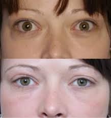
Fractures of the roof of the maxillary sinus with herniation of the orbital content (blow-out fracture), often produce strangulation of the inferior rectus or inferior oblique muscles and thereby impairing movement of eyeball. They can present with periorbital ecchymosis and subconjunctival hemorrhage. Those with fracture of medial wall of orbit without prior surgical intervention can present with enopthalmos. Passage of the endoscope through the intranasal maxillary antrostomy may provide superb visualization of the posterior orbital floor which is the most difficult area to view by transconjunctival or subciliary approach. Anatomically, this area of the orbital floor commonly lies behind the middle half of the globe on axial view. Therefore, the endoscope is an extremely useful means of safely and definitely identifying the posterior edge of the defect and ensuring that the posterior edge of the orbital implant do not compromise the optic nerve or other structures at the orbital apex. In many small fractures for which an implant is not required, endoscopic examination and reduction may be sufficient, thus avoiding an external incisional approach. It is doubtful that endoscopy alone will replace standard CT scanning in the evaluation of orbital fractures. Thus, combining forced duction testing with endoscopic visualization of a fracture may prove to be useful diagnostic tool. Indications include fractures with extension to the middle and posterior portions of the orbital floor, in delayed or secondary fracture repair. Compications includes bleeding, optic nerve damage, medial rectus damage, adhesion, epiphora, periobital emphysema, anosmia, crusting, frontal recess stenosis & osteitis.


